Utah array is a silicon microelectrode array consisting of a grid of sharpened needles etched from a single block of silicon, inserted into the cortex so that each tip records the extracellular voltage near a small number of neurons. Introduced at the University of Utah around 1990, it became and remains the standard sensor for chronic single-neuron recording in humans, and nearly every major human Brain–computer interface result involving individual action potentials has used one.
Its persistence is notable given how little it has changed. A device designed before the field had a single human participant still sets the performance benchmark that newer, higher-channel-count designs are measured against.
How it works
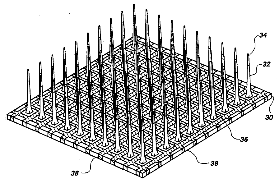
The array is machined from a monolithic block of doped silicon. Dicing saws cut a grid of columns, which are then acid-etched into tapered needles on a 400-micrometre pitch, giving a device roughly four millimetres on a side with 100 electrode sites. The shanks, typically one to one and a half millimetres long, reach the cortical layers where pyramidal-cell somata are dense. The whole structure except the tips is insulated with parylene-C; the exposed tips are coated with platinum or iridium oxide to lower impedance.1
Insertion is the tricky part. Pressed slowly, the array dimples the cortical surface rather than penetrating it, damaging tissue and pial vessels. A pneumatic inserter fires the array in at several metres per second, which produces cleaner penetration.2 Wires from each shank run through a bundle to a connector; in the standard research configuration this is a percutaneous titanium pedestal fixed to the skull, through which amplifiers are plugged in during sessions.
What the array measures is the extracellular field near each tip. Filtered above roughly 250 Hz and thresholded, this yields action potentials from one to a few nearby neurons per channel; filtered below about 300 Hz, it yields local field potentials reflecting activity over a larger volume. Both feed the decoders described under Neural decoding.
Development history
Richard Normann's group designed the array in the late 1980s with a cortical visual prosthesis in mind — a grid of stimulating electrodes in visual cortex intended to evoke a pattern of phosphenes, the same goal that continues to motivate work described under Retinal implants and visual prostheses. The manufacturing process was published in 1991 and the recording characterization followed through the 1990s.
Commercialization passed through several hands: Bionic Technologies, then Cyberkinetics Neurotechnology Systems, then Blackrock Microsystems, now Blackrock Neurotech. Cyberkinetics ran the BrainGate pilot trial, in which Matthew Nagle, paralysed by a knife wound to the spinal cord, received an array in his motor cortex in 2004 and used it to move a cursor, open email, and operate a prosthetic hand.3 The BrainGate2 consortium — Brown, Massachusetts General Hospital, Stanford, and later UC Davis — has run the successor studies since.
In the United States the array holds a clearance for temporary recording of up to thirty days, which supports intraoperative and epilepsy-monitoring use. Every long-term human implant is conducted under an investigational device exemption, not as approved therapy.
Role in human BCI research
Because the array resolves individual neurons, it supports the highest-dimensional control yet demonstrated. Robotic-arm control with multiple degrees of freedom, high-rate cursor typing, and several of the leading Speech neuroprosthesis results rest on Utah arrays, usually two to four implanted across motor and premotor areas; the fastest reported speech rates have come from surface grids rather than penetrating arrays, and the two approaches sit close together. It is also the sensor behind most attempts at writing information back in through intracortical microstimulation, the route by which touch percepts have been delivered to users of Neuroprosthetics.
A variant with shanks of graded length, the Utah slanted electrode array, is designed for peripheral nerve rather than cortex, and has been used to give sensation to users of Myoelectric prosthetics — an early instance of the closed-loop principle that Sensory augmentation research generalizes to senses the body never had.
Depth electrodes of other designs, rather than the Utah array, carry most of the human hippocampal work behind the Memory prosthesis literature, because the relevant structures lie centimetres below the cortical surface that the array is built to sample.
What "96 channels" does and does not meanEach channel may record several neurons whose spikes must be sorted, or none at all. The number of channels yielding usable single units is generally well below the number wired, falls over time, and varies by implant site and participant. Reported channel counts are a property of the hardware, not of the signal obtained.
Failure modes and longevity
Arrays degrade. Some human implants have produced usable neural control for more than a thousand days, and a few for several years, but signal yield generally declines over months to years.4
The causes are mixed. Insertion injury and chronic micromotion provoke a foreign-body response: microglia activate, astrocytes form an encapsulating sheath, and neurons retreat from the tip. Independently, the device itself deteriorates — parylene insulation cracks and delaminates, tip metallization corrodes, wire bonds and connectors fail. A systematic analysis in non-human primates found that material and mechanical failures accounted for a large share of losses, which matters because it implies that improving biocompatibility alone will not fix longevity.5
Percutaneous pedestals add infection risk and prevent the user from living with the system untethered. This is the specific limitation that motivates fully implanted wireless designs, including Neuralink's sealed package and the surface arrays discussed under Electrocorticography interfaces.
Alternatives
Three lines of work aim to displace the array. Higher-density silicon probes, most prominently Neuropixels, put hundreds to thousands of recording sites along a single thin shank and have been used acutely in human cortex during neurosurgery.67 Flexible polymer threads, as used by Neuralink, trade rigidity for reduced chronic tissue reaction but require robotic insertion. Non-penetrating approaches — subdural films and the endovascular Stentrode — give up single-neuron resolution entirely in exchange for stability and lower surgical risk.
A fourth approach avoids metal electrodes altogether: Optogenetics can read and write neural activity optically, but requires genetically modifying the target neurons, which places it far from routine human use.
None has yet accumulated a comparable human track record. That is the array's main remaining advantage: two decades of implants across multiple sites, with known failure statistics, against competitors whose chronic behaviour in humans is largely unmeasured.
Outlook
The realistic future for penetrating arrays is not that they win or lose outright but that they specialize. Applications demanding many independent control dimensions — dexterous limb control, fluent speech, and any bidirectional system delivering fine sensory feedback — need single-neuron resolution that surface and vascular electrodes cannot supply. Applications needing a reliable switch do not.
Whether that specialization survives depends on an unresolved empirical question: whether a penetrating device can be built that still records well after ten years in a human brain. The best-established implanted neurotechnologies, among them the Cochlear implant and Deep brain stimulation, stimulate rather than record and sit in tissue that tolerates them for decades. No recording array has demonstrated equivalent durability, and the failure analyses suggest the answer depends as much on materials engineering as on neuroscience.
See also
- Brain–computer interface
- Neural decoding
- Speech neuroprosthesis
- Electrocorticography interfaces
- Stentrode
- Neuralink
- Neuroprosthetics
- Memory prosthesis
References
Footnotes
-
paperCampbell, P. K., Jones, K. E., Huber, R. J., Horch, K. W. and Normann, R. A. "A silicon-based, three-dimensional neural interface: manufacturing processes for an intracortical electrode array." IEEE Transactions on Biomedical Engineering, 1991. ↩
-
paperMaynard, E. M., Nordhausen, C. T. and Normann, R. A. "The Utah intracortical electrode array: a recording structure for potential brain-computer interfaces." Electroencephalography and Clinical Neurophysiology, 1997. ↩
-
paperHochberg, L. R. et al. "Neuronal ensemble control of prosthetic devices by a human with tetraplegia." Nature, 2006.↩Centres on the first BrainGate participant, performing tasks in supervised laboratory sessions rather than in independent daily use.
-
paperSimeral, J. D., Kim, S.-P., Black, M. J., Donoghue, J. P. and Hochberg, L. R. "Neural control of cursor trajectory and click by a human with tetraplegia 1000 days after implant of an intracortical microelectrode array." Journal of Neural Engineering, 2011.↩One participant and one long-lived implant; it establishes that the duration is possible, not that it is typical.
-
paperBarrese, J. C. et al. "Failure mode analysis of silicon-based intracortical microelectrode arrays in non-human primates." Journal of Neural Engineering, 2013. ↩
-
paperJun, J. J. et al. "Fully integrated silicon probes for high-density recording of neural activity." Nature, 2017. ↩
-
paperPaulk, A. C. et al. "Large-scale neural recordings with single neuron resolution using Neuropixels probes in human cortex." Nature Neuroscience, 2022.↩Recordings were made acutely during neurosurgery; these probes have no chronic human track record to compare with the array's.