Loading
Old version — revision 1
This is a fixed snapshot of Organ bioprinting, saved by Import as part of the initial corpus import. It is not edited and it is not updated; the article may have changed since.
Edit summary: Initial import of content/organ-bioprinting.md — the filesystem corpus, unchanged. Not an edit.
Organ bioprinting
The additive manufacture of living tissue by depositing cells and biomaterials in a programmed pattern, pursued as a route to transplantable organs but limited so far to simple structures.
TechnologyExperimental2040sTRL 4revision 1
16.6 kB1,742 wordsBodiesfrontmatter date 2026-07-27
Organ bioprinting is the additive manufacture of living tissue: a machine deposits cells, hydrogels and sacrificial support materials in a programmed three-dimensional pattern, and the construct is then cultured until the cells remodel it into something with tissue-like structure and function. The technique borrows its motion control from industrial 3D printing and its biology from Tissue engineering. It has produced skin, cartilage, corneal stroma, vascular grafts and beating cardiac patches in the laboratory. It has not produced a solid organ that anyone has transplanted into a person, and the gap between those two statements is the subject of this article.
~200 µm
Maximum viable distance from a capillary
beyond which cells outstrip passive oxygen diffusion
10⁸/mL
Cell density of native solid tissue
orders of magnitude above most printable bioinks
0
Printed solid organs implanted in humans
as of 2026
How it works
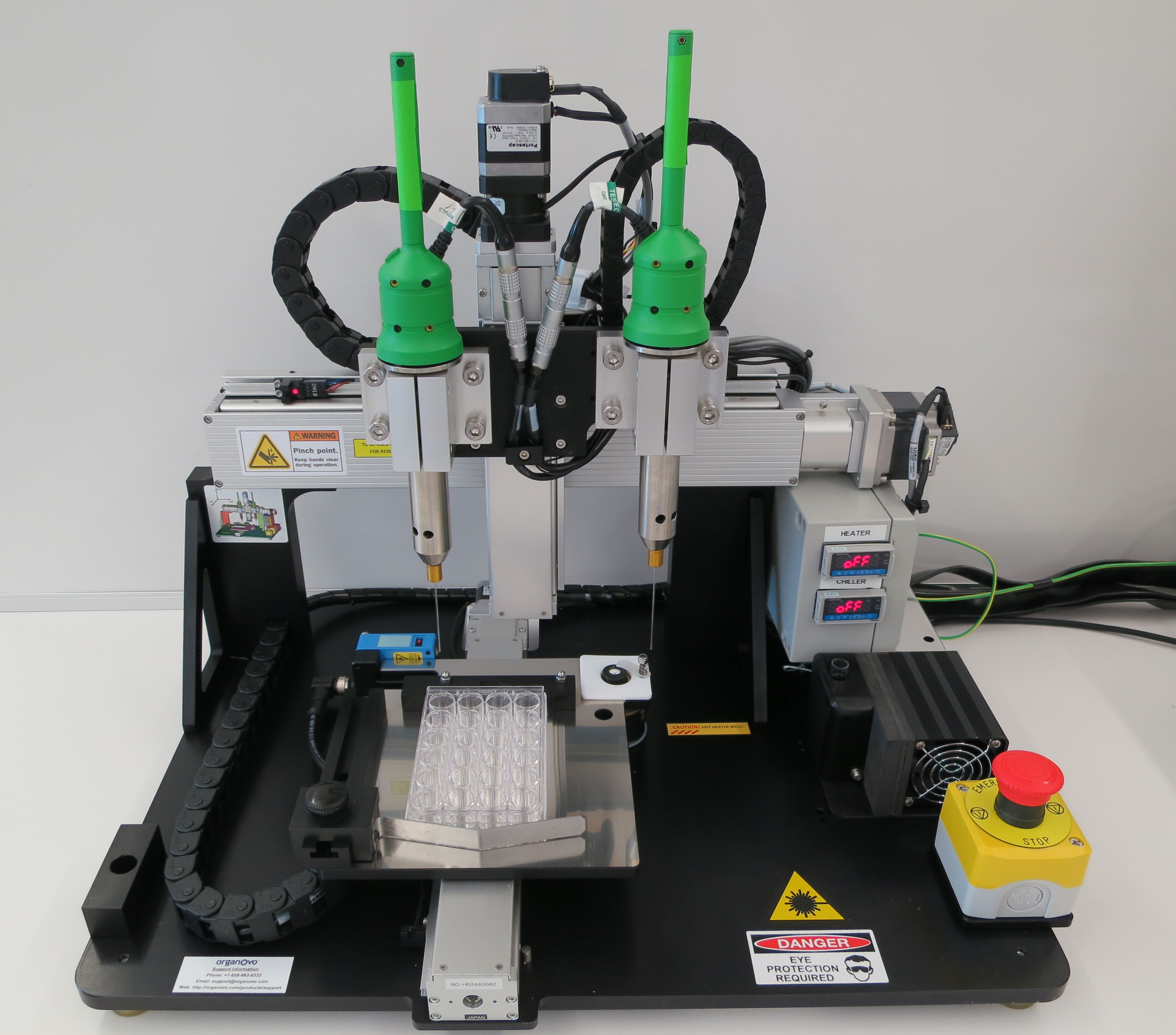
A bioprinter converts a digital model, usually derived from a patient's CT or MRI scan, into a toolpath and then lays down material along it. Three deposition families dominate.
Extrusion printing pushes a viscous cell-laden gel through a nozzle by pneumatic pressure or a screw. It is the workhorse: cheap, tolerant of high cell densities, compatible with most hydrogels. Its weaknesses are resolution, typically a few hundred micrometres, and shear stress at the nozzle, which damages cells. Extrusion also cannot print an unsupported soft gel — a low-stiffness ink collapses under its own weight. The standard answer is embedded printing, in which the nozzle deposits into a supporting bath of granular gel that behaves as a solid at rest and yields locally around the moving needle. The FRESH method developed in Adam Feinberg's laboratory uses a gelatin microparticle slurry for this and was used to print collagen replicas of heart valves and a neonatal-scale ventricle that contracted when seeded with cardiomyocytes.1
Droplet and inkjet printing eject picolitre droplets of low-viscosity cell suspension. Resolution is better and cell damage lower, but the constructs are mechanically weak and the technique struggles with anything thick.
Light-based printing cures a photosensitive resin with patterned light. Digital light processing projects an entire layer at once, which is fast and precise; two-photon polymerization reaches sub-micrometre features but is far too slow for organ-scale volumes. Volumetric printing, in which tomographic light projections are rotated around a resin vial so that a whole object solidifies at once where the accumulated dose crosses a threshold, produces centimetre-scale constructs in tens of seconds and avoids layering artefacts altogether.2 A landmark demonstration used food-colouring dyes as biocompatible photoabsorbers to print hydrogels containing intertwined, topologically independent vascular networks, including an air-sac model that oxygenated human red blood cells flowing past it during cyclic ventilation.3
Bioinks
A bioink must satisfy contradictory requirements. It has to flow through a nozzle or cure under light, hold its shape afterwards, stay soft enough for cells to remodel, degrade at the rate new matrix is deposited, and be non-toxic throughout. Common bases are alginate, gelatin methacryloyl, fibrin, collagen I, hyaluronic acid, and decellularized extracellular matrix digested from donor tissue, which brings tissue-specific composition at the cost of batch variability. See Decellularized scaffolds for that material family.
The deeper problem is cell density. Native solid tissue packs cells at roughly a hundred million per millilitre; a printable gel usually carries one to two orders of magnitude fewer, because at high density the ink loses printability and the cells lose oxygen. One workaround inverts the usual arrangement: rather than printing cells in a gel, the SWIFT approach compacts organoid-derived cells into a dense living matrix and prints only the sacrificial vascular network into it, then washes the sacrificial ink out to leave perfusable channels.4 The tissue is the bulk; the print is the plumbing.
Development history
-
1988Cells through a printerRobert Klebe adapts an office inkjet to position cells on a substrate, coining 'cytoscribing' and establishing that printing hardware need not kill cells.
-
2003Thermal inkjet bioprintingThomas Boland's group at Clemson shows that modified commercial inkjet printers can deposit viable mammalian cells in patterns.
-
2012Sacrificial vasculatureJordan Miller and colleagues print lattices of carbohydrate glass, cast tissue around them and dissolve them away, leaving open perfusable channels inside a cell-laden gel.
-
2019Three converging advancesEmbedded collagen printing produces heart components; tomographic light projection prints centimetre-scale objects in seconds; entangled multivascular networks are printed in a single monolithic gel.
-
2022First printed implant in a trial3DBio Therapeutics implants an ear reconstructed from a patient's own chondrocytes in a microtia clinical trial, the first bioprinted living implant placed in a person under regulatory oversight.
-
2023–2026Industrialization without organsBioprinted tissue moves into drug screening and into partnerships with pharmaceutical firms for implantable therapeutic tissues; no solid organ reaches the clinic.
What has actually been implanted
The honest inventory is short and consists of tissues that are thin, avascular, or both.
Printed cartilage has reached patients. An ear scaffold printed from a patient's expanded chondrocytes was implanted in a microtia trial beginning in 2022; cartilage is a natural first target because it is avascular, mechanically simple and immunologically quiet when autologous. Printed skin substitutes have been tested in animals and in early human work, including handheld and robot-mounted devices that deposit cells directly onto a wound bed. Printed bone-graft substitutes and airway splints exist, but most of these are acellular polymer devices produced by conventional 3D printing rather than bioprinting proper, and the distinction matters: a resorbable printed splint is a device, while a construct containing living cells is regulated as a biologic or a combination product.
Bioprinting hardware has also been flown to the International Space Station, on the reasoning that in microgravity a soft construct holds its shape without a support bath. The samples produced there are small, and the motivation overlaps with the broader interest in manufacturing medical supplies far from resupply described in Space medicine.
Everything organ-shaped remains a laboratory object. A widely reported 2019 construct from Tal Dvir's group at Tel Aviv University, printed from a patient's cells in a bioink derived from their own omentum, was about the size of a cherry, had the gross anatomy of a heart, and did not pump.5 The FRESH collagen ventricle contracted but produced negligible pressure. No printed kidney, liver or heart has supported an animal, let alone a person. Where whole-organ replacement has actually reached patients, it has done so through Xenotransplantation or through mechanical substitutes such as the Artificial heart, not through printing; the competing strategy of growing an organ from cells rather than depositing it is treated in Lab-grown organs.
What "printed a heart" meansPress coverage of bioprinting routinely reports the geometry as though it were the achievement. Printing the shape of an organ is a solved problem; a desktop machine can do it in a day. Producing tissue with the correct cell types, in the correct densities, arranged around a perfused vascular tree, at a mechanical and metabolic standard that keeps a human alive, is unsolved and not close to solved.
The vascularization bottleneck
Every cell in a solid organ sits within roughly a hundred to two hundred micrometres of a capillary, because that is how far oxygen diffuses through tissue before consumption exhausts it. Any construct thicker than about a millimetre without internal perfusion develops a necrotic core within days. This single constraint explains the field's shape: the tissues that work clinically are sheets and small pieces, and the tissues that do not work are everything else.
Printed vascular networks solve part of the problem and expose the rest. Current printers resolve channels down to a few hundred micrometres reliably and to tens of micrometres in specialised light-based systems. Human capillaries are five to ten micrometres across, branch across roughly a dozen generations, and in a kidney or liver form a network whose total surface area is measured in tens of square metres. No printing process approaches that resolution at organ scale in an acceptable time. The prevailing strategy is therefore hybrid: print the large and medium vessels, seed them with endothelial cells, and rely on angiogenic sprouting from those vessels to build the capillary bed biologically. Whether self-assembly can reliably complete a network that a printer starts, and do so before the tissue starves, is the central open question.
Perfusion also has to be surgically connectable. A printed construct needs an inlet and an outlet that a surgeon can anastomose to the recipient's circulation, with wall strength sufficient to hold arterial pressure and an endothelial lining continuous enough not to trigger clotting. An incompletely endothelialized channel thromboses, which is the same failure mode that defeated recellularized donor scaffolds and that limits Artificial blood and mechanical circulatory support.
Limitations and risks
Cell supply. A human-scale solid organ contains cells on the order of ten to a hundred billion. Sourcing them autologously from Induced pluripotent stem cells means months of expansion and differentiation per patient at high cost, on a timescale incompatible with acute organ failure. Allogeneic cells shorten the timeline but reintroduce immunosuppression, which is much of what a grown organ was supposed to avoid. Where the organ failed because of an inherited defect, the patient's own cells carry that defect and must be corrected before printing, adding the manufacturing burden of Somatic gene therapy to the tissue problem.
Maturation. Printed tissue starts fetal-like. Cardiomyocytes derived from stem cells contract weakly and lack adult calcium handling; hepatocytes lose function within days in culture. Maturation requires mechanical and electrical conditioning in bioreactors over weeks, and it is incomplete. The same immaturity ceiling constrains Organoids.
Quality control and regulation. A printed organ is a patient-specific manufactured product, which sits awkwardly in regulatory frameworks built either for mass-produced devices or for standardized biologics. Regulators have issued guidance on additive manufacturing of devices while explicitly leaving cell-containing products outside its scope. Sterility, potency assays, batch release testing and shelf life all have to be defined for a product with a batch size of one.
Tumorigenicity. Constructs built from pluripotent-derived cells carry residual risk of undifferentiated cells forming teratomas, the same hazard that constrains Epigenetic reprogramming and stem-cell therapies generally.
Cost and equity. If a printed organ works, it would initially be expensive and produced by a small number of facilities. The distribution problem that follows is not technical, and Access and inequality applies to it in full.
Measured on the scale set out in Technology readiness level, the field spans an unusual range: printed skin and cartilage sit near clinical validation, printed vascularized organs remain at proof of concept, and the two are routinely reported under the same heading.
Outlook
The near-term commercial reality of bioprinting is not transplantation but tissue models: printed liver, kidney and tumour tissue used for toxicology and drug screening, a market that grew as regulators softened the requirement for animal testing in some drug categories. Therapeutic products under active development are small and encapsulated — printed islet constructs for type 1 diabetes, printed cartilage, printed nerve conduits, printed corneal tissue — where a few cubic centimetres of tissue can do clinical work without a vascular tree.
For solid organs, several groups now argue that printing will not be the fabrication method at all, but the scaffolding method: printing the vasculature and letting cells build the parenchyma. That reframing makes the problem tractable in principle and leaves it hard in practice, and it borrows its logic from developmental biology rather than manufacturing — the same logic that makes the study of animals capable of Limb regeneration relevant to a discipline built around machine tools. A useful benchmark for the field's honesty is that no printed construct has yet supported a large animal's physiology for a month. Until one does, timelines for a printed kidney rest on extrapolation rather than evidence, and the Organ shortage that motivates the whole enterprise continues to be addressed by donation reform, machine perfusion and edited pigs.
See also
- Tissue engineering
- Decellularized scaffolds
- Lab-grown organs
- Organoids
- Organ shortage
- Xenotransplantation
- Artificial heart
- Induced pluripotent stem cells
References
Footnotes
-
paperLee, A. et al. "3D bioprinting of collagen to rebuild components of the human heart." Science, 2019. ↩
-
paperBernal, P. N. et al. "Volumetric bioprinting of complex living tissue constructs within seconds." Advanced Materials, 2019. ↩
-
paperGrigoryan, B. et al. "Multivascular networks and functional intravascular topologies within biocompatible hydrogels." Science, 2019.↩The oxygen-transfer demonstration used a hydrogel air-sac model perfused on the bench; it is a materials and geometry result, not living lung tissue.
-
paperSkylar-Scott, M. A. et al. "Biomanufacturing of organ-specific tissues with high cellular density and embedded vascular channels." Science Advances, 2019. ↩
-
paperNoor, N. et al. "3D printing of personalized thick and perfusable cardiac patches and hearts." Advanced Science, 2019.↩Reported worldwide as the first 3D-printed heart; the heart-shaped construct was cherry-sized and did not pump, and the contribution is the patient-derived bioink.